After the decision to treat the breast with radiation therapy has been made, the next step is to decide which areas of the breast are at risk and the dosage.
There are several machines and techniques available to deliver the treatment. Most of the times doctors will end up choosing between a 3DCRT based treatment or a VMAT based treatment.

In both treatment plans a CT scan will be performed in a position that can be replicated during treatment.
Another similarity is that the volumes of radiation will be adapted to your anatomy and both techniques are equal in terms of disease control and overall survival.
In this type of treatment, the machine is positioned in a way the radiation oncologist calls tandem beams. In this configuration there will be a direct beam of radiation that will cover the area in a straight line.
The main advantage is that outside of this straight line there is very little radiation that will reach other organs, this leads to less fatigue, esophagitis and nausea.
The main disadvantage is that organs that lie inside this straight line will receive as much radiation as the main area of the breast, so important organs may receive the full dose if they are close to the area at risk.
This may also translate in certain types of side effects not seen in VMAT planning. This also leads to more skin reaction and implant related complications.
This treatment variation involves rotating a beam moving around the body in an arc to target areas deemed at risk by the physician.

The advantage is that the treatment machine can focus on specific areas while sparing others, especially if the radiation treatment volume is close to a sensitive organ that should be avoided.
This approach may be preferred in patients with certain lung anatomy that may require circling around the lungs or if the tumor is near the sternum.
By selecting which areas to focus on, the technique has demonstrated a reduction in reconstruction failure rates.
However, a disadvantage is that a larger volume of the chest receives a low dose of radiation, leading to potential long-term side effects such as an increased risk of heart damage and the development of a second cancer.
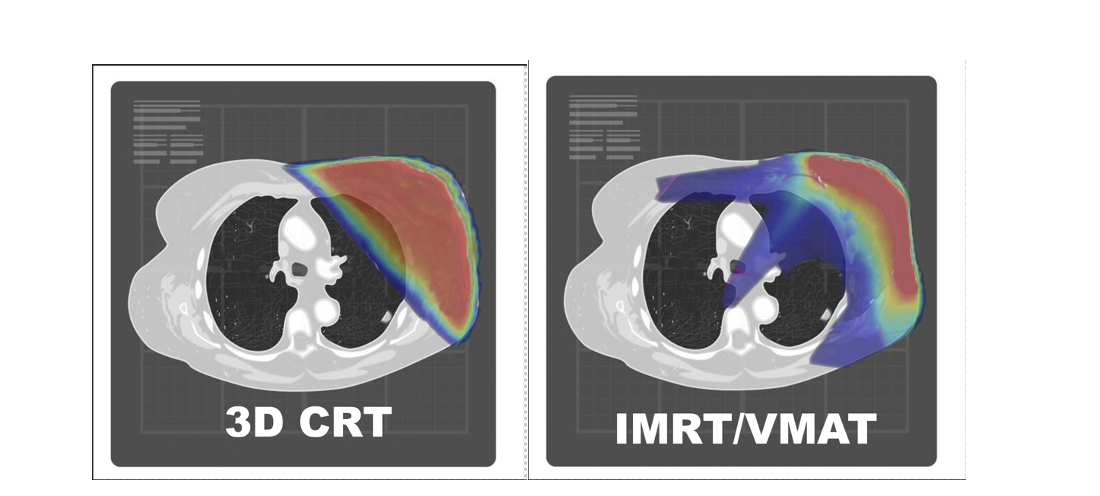
The high area doses are the redder zones, while the blue ones are areas that receive low dose of radiation.
Notice how in 3DCRT there is a slightly greater volume that receives a high dose and in IMRT there is a big volume that receives a low dose.
|
Advantage 3DCRT |
Advantages IMRT |
| Decreased risk of secondary malignancies |
Might reduce heart dose in certain cases |
|
Less gastrointestinal symptoms |
Less time to perform |
|
Less fatigue |
Less skin reaction |
|
Less esophagitis |
Less implant reconstruction failure |
|
|
In advanced cases may allow contouring certain hard to get areas |
|
Same |
|
|
Chances of cancer control |
|
|
Need for a CT scan to plan treatment |
|
|
Number of sessions needed to complete treatment |
|